Friday, December 23, 2011

Gene Therapy in Ophthalmology Update 6: First-Ever Clinical Trial for the Autosomal Recessive Form of Retinitis Pigmentosa (arRP) is Underway

It has been difficult keeping up with the changing world of gene therapy in ophthalmology, but thanks to the Foundation Fighting Blindness, I learned yesterday about this new, and first clinical study for treating a rare form of retinitis pigmentosa, underway in Saudi Arabia.

Here is the story, as reported by the FFB’s website:


First Gene Therapy Clinical Trial for Recessive RP is Underway

December 22, 2011 - The field of gene therapy for retinal degenerative diseases is taking a big step forward with the launch of the first-ever clinical trial for people with an autosomal recessive form of retinitis pigmentosa (arRP). The human study, underway at King Khaled Eye Specialist Hospital in Riyadh, Saudi Arabia, is evaluating gene replacement for individuals with mutations in the gene MERTK, which is a frequent cause of arRP in people of Middle Eastern descent.

While the primary goal of the six-participant, Phase I trial is to evaluate the treatment's safety, investigators will also be looking at its effect on vision.

The treatment works by using a manmade adeno-associated virus, or AAV, to deliver healthy copies of the MERTK gene to cells in the retina. The treatment is contained in a tiny drop of liquid that is injected underneath the retina and absorbed by a layer of cells called the retinal pigment epithelium (RPE).

The MERTK gene plays an important role in the daily maintenance and regeneration of photoreceptors, the retinal cells that enable people to see. During sleep, the tips of photoreceptors are shed and disposed of by the RPE through a process called phagocytosis. Subsequently, the tips grow back. However, when the MERTK gene is defective, the disposal and regeneration process doesn't work properly, and debris and waste products accumulate, causing photoreceptor death and vision loss

"It is great to see clinical trials of gene therapy expanding into more forms of retinal disease and targeting additional mechanisms of disease such as defects in phagocytosis," says Stephen Rose, Ph.D., chief research officer, Foundation Fighting Blindness. "If successful, it broadens the range of retinal conditions that are amenable to gene therapy."

Dr. Rose notes that, for years, the Foundation has funded several MERTK and phagocytosis research projects, which helped make the current study possible. In addition, the AAV being used for gene delivery is similar to the one used in clinical trials of gene therapy that have restored vision in children and young adults virtually blind from Leber congenital amaurosis.

The MERTK gene therapy trial is being led by Drs. Kang Zhang of the University of California, San Diego, and Fowzan Alkuraya of King Khaled Eye Specialist Hospital. Dr. William Hauswirth, a Foundation-funded gene therapy development expert from the University of Florida, is also on the study team. His lab developed the AAV used in the trial and carried out preclinical safety studies to gain approval for the trial in Saudi Arabia.


Editor’s Note: I have been trying to keep track of the many pre-clinical and clinical studies underway in this ever changing field. I have put together a table of all of the activities underway that I have been able to identify and offer it to all interested parties. Gene Therapy Companies/Institutions Active in Ophthalmology, Version 8, updated as of yesterday, is available in a Word version to any that request it. Use the Email Me! link shown in the right-hand column.

Thursday, December 01, 2011

Gene Therapy in Ophthalmology Update 5: A Complement-Based Gene Therapy for AMD

Selected Reviews of AAO 2011 Retina SubSpecialty Day Presentations

Here is another overview of a presentation made during the Retina SubSpecialty Day Meeting. 

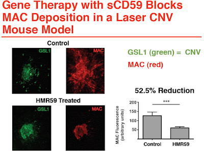
Dr. Elias Reichel, of Tufts University School of Medicine and a founder of Hemera Biosciences, Inc., of Boston, MA, presented on a new approach to treating the dry form of age-related macula degeneration. His paper was based on the research being done by Hemera Biosciences on HMR59, a naturally occurring protein that protects retinal cells from damage by MAC (Membrane Attack Complex), that can be delivered for long-lasting activity via a gene therapy approach. 

HMR59 was developed at Tufts University and subsequently licensed to Hermera Biosciences.  


Complement Regulation via Gene Therapy for Dry AMD 

Elias Reichel, M.D., Professor of Ophthalmology, Tufts University School of Medicine, Boston, MA

HMR59 is a novel therapy primarily targeted at geographic atrophy and other forms of dry age related macular degeneration (Dry AMD) by blocking the final stage of the complement cascade, membrane attack complex (MAC). The complement cascade is implicated via genetic studies as playing a critical role in both wet and dry AMD. 


HMR59 is a gene therapy using an AAV2 vector to express a soluble form of a naturally occurring membrane bound protein called CD59 (sCD59), which blocks MAC. Membrane attack complex is the final common pathway of activation of the complement cascade, and is composed of complement factors C5b, C6, C7, C8 and C9 that assemble as a pore on cell membranes. The MAC pore induces ionic fluid shifts leading to cell destruction and ultimate death. 

HMR59 works by increasing the production of sCD59 by ocular cells. The sCD59 released from the cells will circulate throughout the eye and penetrate the retina to block MAC deposition and prevent cellular destruction. By blocking MAC, the remainder of the upstream complement cascade is left intact to perform its normal homeostatic roles. 


HMR59 will be injected directly into the vitreous cavity in an office setting. Such a procedure is currently performed by all retinal specialists using other medications quite commonly. Using the gene therapy approach offers the opportunity to reduce the number of injections needed over a patient's lifetime as the ocular cells will act as factories to produce sCD59, thus addressing the issue of drug delivery. 


In summary: 

• The complement pathway is strongly associated with AMD 
• Membrane attack complex (MAC) is the final step in the complement pathway 
• CD59, a naturally occurring protein, protects retinal cells from damage by MAC 
• Hemera Biosciences has developed a gene therapy which produces soluble CD59 (sCD59) that blocks MAC 
• A single intravitreal injection allows for long term protection from AMD progression 


Hemera Biosciences is currently seeking funding to begin animal toxicology studies to get to a phase 1 study in humans. 

Inquiries for further information should be made to: Elias Reichel, MD


Wednesday, November 16, 2011

Gene Therapy in Ophthalmology Update 4: Table of Companies and Institutions Participating

As a followup to the stem cells in ophthalmology table recently published, here is one tabulating the participants using gene therapy in ophthalmology.

When I first wrote about gene therapy in ophthalmology (Gene Therapy for RP and Dry AMD) in November 2010, I began collecting information about the various participants with the thought of writing a Primer, similar to the one I did on stem cells in ophthalmology. At that time, I was able to identify seven companies participating. Since then, and with the help of a few friends, I have now identified nearly thirty companies and institutions using gene therapy approaches to treat ophthalmic diseases. Since I believe access to knowledge is very important, here is the latest version of my table of companies and institutions using gene therapy approaches for treating ophthalmic diseases.

Please let me know of any corrections or omissions.


(An easier to read pdf file of this table is available from the author via email request.)

Saturday, November 12, 2011

Stem Cells in Ophthalmology Update 12: Updated Table of Company Participants

When I first wrote about stem cells in ophthalmology (Primer) in September 2010, I was able to identify six companies participating. Since then, and with the help of a few friends, I can now identify eleven companies using stem cells to treat ophthalmic diseases. Since I believe access to knowledge is very important, here is my revised table of companies using stem cells in treating ophthalmic diseases.

Please let me know of any corrections or omissions.


(An easier to read pdf file of this table is available from the author via email request.)

Friday, November 11, 2011

Iluvien Update 4: FDA Turns Down Alimera’s NDA for Approval of Iluvien Again

Alimera again received bad news from the FDA on its application for approval of Iluvien. In a letter (a CRL or complete response letter), the FDA said that questions remained based on the data previously submitted, about the adverse reactions shown by Iluvien in the FAME Study (risk of cataracts and raised IOPs) and that these were not offset by the benefits demonstrated.

The FDA indicated that Alimera would need to conduct two additional clinical trials to demonstrate that the product is safe and effective for the proposed indication.

Here is Alimera’s complete new release:


Alimera Sciences Receives Complete Response Letter From FDA for ILUVIEN(R)
Conference Call Scheduled for Monday, November 14 at 8 a.m. Eastern Time

ATLANTA, Nov 11, 2011 (GlobeNewswire via COMTEX) -- Alimera Sciences, Inc., ("Alimera"), a biopharmaceutical company that specializes in the research, development and commercialization of prescription ophthalmic pharmaceuticals, today announced that it has received a complete response letter (CRL) from the U.S. Food and Drug Administration (FDA) in response to the New Drug Application (NDA) for ILUVIEN(R) for the treatment of diabetic macular edema (DME) associated with diabetic retinopathy.

A CRL is issued by the FDA's Center for Drug Evaluation and Research when their review of an application is completed and questions remain that precludes the approval of the NDA in its current form.

The FDA stated that it was unable to approve the ILUVIEN NDA because the NDA did not provide sufficient data to support that ILUVIEN is safe and effective in the treatment of patients with DME. The FDA stated that the risks of adverse reactions shown for ILUVIEN in the FAME(R) Study were significant and were not offset by the benefits demonstrated by ILUVIEN in these clinical trials. The FDA has indicated that Alimera will need to conduct two additional clinical trials to demonstrate that the product is safe and effective for the proposed indication.

The Company will be requesting a meeting with the FDA to clarify next steps.

ILUVIEN is Alimera's investigational, sustained drug delivery system that releases sub-microgram levels of fluocinolone acetonide (FAc) for the treatment of DME. In December 2010, the FDA issued a CRL to Alimera related to its June 2010 NDA for ILUVIEN, which included data through month 24 of the FAME(TM) Study. In that first CRL, the FDA asked for, among other things, analyses of the safety and efficacy data through month 36 of the FAME Study. Alimera submitted a response to the FDA on May 12, 2011, addressing the issues raised in the first CRL and including 36-month trial data. The FDA classified Alimera's response as a Class 2 resubmission, resulting in a six-month review period and a Prescription Drug User Fee Act, or PDUFA, date of November 12, 2011.

"We are surprised and disappointed with the FDA's decision on our application to market ILUVIEN in the U.S. to patients with this devastating disease. Based on extensive research with U.S. retinal physicians, we have learned that ILUVIEN's long-term sustained delivery treatment benefit is desired and that ILUVIEN has a manageable risk to benefit ratio. We continue to believe in ILUVIEN as a long-term effective treatment option for DME. We are committed to, and have the funds for, pursuing approval in Europe and for evaluating our options in the U.S.," said Dan Myers, president and chief executive officer of Alimera.

For Europe, Alimera expects to submit its formal response to the Preliminary Assessment Report to the Medicines and Healthcare products Regulatory Agency (MHRA) later this month. Based on this submission, the MHRA is expected to make a recommendation on the approvability of ILUVIEN to Alimera and the Concerned Member States (Austria, France, Germany, Italy, Portugal and Spain) by the end of this year, with a decision regarding the approval of ILUVIEN expected in the first half of 2012. The market opportunity in Europe is similar in size to the U.S. market opportunity.

Wednesday, November 09, 2011

Fighting Retinal Disease: The Promise of New Approaches

Dr. Stephen Rose, the Chief Research Officer of the Foundation Fighting Blindness has just written an article about emerging treatments for retinal diseases, including the use of stem cells, gene therapy and new pharmaceutical approaches. I feel it is important for those of you on the front lines fighting these diseases and those of you who have them, to know that we are getting closer and closer to solving some of the questions about saving sight, and perhaps even bringing back lost sight.

Please take a look at Dr. Rose’s words and dig deeper into the resources available on the FFB website. (I have not included the links that were included with some of his remarks, but you can go to the original on the web and check them out for yourselves.)


What Emerging Treatment Will Work Best for Me?


Written by Stephen Rose, Ph.D., Chief Research Officer, Foundation Fighting Blindness


November 8, 2011 - Hope has never run higher in the fight against retinal degenerative diseases, thanks to clinical trials now underway for gene, stem cell and pharmaceutical therapies. As a result, people affected by these vision-robbing conditions are naturally eager to figure out what emerging treatment approach is going to work best for them.

While it is tempting for someone affected to focus on a "magic bullet" to stop or reverse their disease, I strongly encourage people to consider the Foundation's comprehensive portfolio of emerging treatments when thinking about the future of their vision. Here are three important reasons why:

We can't predict how future research will unfold

Science always has surprises. As an example, until a year or two ago, we believed that corrective gene therapy would be suitable only for treating early-stage disease when a person had a lot of photoreceptors left to save. But recently, Foundation-funded researchers from the University of California, Berkeley and other groups are using gene therapy to enable ganglion cells -- cells in the retina that survive long after photoreceptors are lost -- to provide vision. This research advance has now put gene therapy on the map for potentially reversing total blindness. Previously, we thought that only stem cells or artificial retinas could restore vision in advanced disease.

We need to have back-up plans

In the next couple of years, as we see a big increase in the number of clinical trials for inherited retinal diseases, some setbacks will be inevitable. We need to keep in mind that clinical trials are experiments and, invariably, will not always achieve optimal results. It is critical that we have multiple treatments available, or in the pipeline, for each disease, in case a promising course of action does not pan out. This is one reason pharmaceutical therapies are so important. Generally speaking, they can treat a broader range of conditions than, for example, a gene therapy directed at a specific genetic defect. I envision pharmaceuticals often serving as bridges, or as alternatives, to more targeted therapies. 

Treatment decisions will be personal

Several factors will play a role in determining which treatments might work best for an individual, including his or her genetic profile, stage of disease, age and even tolerance for risk. Let's say in the future that a middle-aged person with retinitis pigmentosa has been taking a drug for many years that's done a good job preserving vision, and along comes a new gene or stem cell therapy. Does that person try a new treatment or stick with what is known to work? There is no right or wrong course of action. What's important is that we fund a diversified portfolio of research, so patients have options. Multiple treatment alternatives may seem like a luxury now, but we are working hard to make that day a reality as soon as possible.

Find out more about Foundation research

The Foundation currently funds 130 grants at 73 institutions around the world. Grants are selected through a rigorous review process conducted by the Foundation's Scientific Advisory Board, which is comprised of the world's top retinal researchers. A complete list of the Foundation's grants is at: www.FightBlindness.org/grants.

Wednesday, November 02, 2011

Gene Therapy in Ophthalmology Update 3: Genetic Testing of RP Patients Necessary in Order to Direct Treatment

Selected Reviews of AAO 2011 Retina SubSpecialty Day Presentations
Here is another of the presentations made during the Retina SubSpecialty Day Meeting.

Dr. Stephen Tsang presented on factors and the genetics of retinitis pigmentosa. His paper was based on the article previously published by he and his co-author, Kyle Wolpert, that appeared in the November 2010 issue of Retinal Physician
.

The Genetics of Retinitis Pigmentosa
Knowing how the varieties of RP are transmitted can be half the battle of treatment.

Stephen H. Tsang, MD, PhD and Kyle Wolpert, BA

Published in Retinal Physician, November 2010
(Reprinted with permission of the authors)

Retinitis pigmentosa (RP) is a heterogeneous group of diseases characterized by progressive rod-cone dysfunction. Patients initially present with nyctalopia from rod photoreceptor loss, progress to tunnel vision and ultimately experience central vision loss. RP is also the most common form of inherited retinal degeneration, affecting one in 3,000 people.(1,2)

GENETICS BASICS

As a genetically heterogeneous set of disorders, the specific mutation involved in any given case of RP dictates the inheritance pattern and strongly influences the prognosis. As such, a careful family history is essential both for diagnosis and genetic counseling. When possible, the family members of a new RP patient should be examined in order to better define the inheritance pattern. Often, family members may be younger than the age at which the disease develops, which can make this process difficult.

Electroretinogram (ERG) testing, which provides a global assessment of rod and cone function, can measure electro-physiological disturbances long before photoreceptor loss occurs or changes can be seen on fundus examination.(1,3) Thus, ERG testing can help determine whether younger family members will present with the disease later in life, which is useful both for the construction of a pedigree and also for counseling purposes; for example, ERG screening may help a young patient identify plausible career paths.

Described inheritance patterns of RP include autosomal dominant (15% to 35% of cases), autosomal recessive (60%), X-linked recessive (5% to 18%), and mitochondrial. If no other family members are affected, the disease is likely the result of an autosomal recessive (AR) mutation. If the disease presents only in men and is transmitted maternally, then it is likely an X-linked recessive (XLR) mutation. Unlike the recessive modes of inheritance, autosomal dominant (AD) transmission is marked by disease occurrence in every generation and father-to-son transmission.

The rarest form of inheritance is X-linked dominance. These patients are almost always women, as such traits are generally lethal in men. It is possible that a sporadic case could represent a new autosomal dominant mutation, but this is rare. However, the possibility underscores the need for genetic testing to ensure an accurate diagnosis. Mitochondrial mutations are passed maternally and often present systemic problems. Identifying the inheritance pattern involved can help to determine the prognosis, both for the patient and the rest of his or her family.

AUTOSOMAL DOMINANT

Between 15% and 35% of all cases of RP follow an autosomal dominant inheritance pattern.(4,5) As stated previously, AD inheritance is marked by occurrence in each generation and father-to-son transmission of the disease. Compared to AR forms, the AD forms of the disease tend to be more mild, progress more slowly, and present later in life. Patients present with reduced visual acuity and loss of color vision in late adulthood and progress to legal blindness. In the first two decades of life, patients with autosomal dominant RP may be funduscopically indistinguishable from healthy patients. Mutations in rhodopsin, the visual pigment, are responsible for 30% of AD forms of RP. In patients with RP, autofluorescence imaging often shows a characteristic ring of hyperautofluorescence before abnormalities appear on fundus examination; as such, autofluorescence imaging can help to provide presymptomatic clinical evaluation of the patient and predict the course of the disease.(6-8)

Genetic counseling for patients with AD RP is relatively straightforward. Assuming that only one of the patient's parents is affected, each of the patient's children will have a 50% chance of inheriting the mutant allele and thus the disease. Furthermore, an affected patient's siblings each have a 50% chance of being affected. Siblings and children that do not have the allele (as determined by ERG testing) will not pass the disease on to their own children.

AUTOSOMAL RECESSIVE

Autosomal recessive forms of retinal degeneration tend to be more severe, progress more rapidly, and present earlier than the AD forms. As stated previously, the AR inheritance pattern is characterized by sporadic appearance and occurrence in both men and women.

Genetic counseling for patients with AR retinitis pigmentosa is more complicated than for the AD forms. If a patient is affected with an AR form of RP, then both of their parents must have been heterozygous carriers. This means that each of their siblings has a 25% chance of developing the disease and a 50% chance that they are asymptomatic carriers; thus, if a sibling does not have the disease, then there is still a 66% chance that they are carriers. The patient's children will not develop the disease, but each will be a heterozygous carrier, so it may reappear in later generations.

X-LINKED RECESSIVE

The X-linked recessive form of retinal degeneration is often the most severe. It has an early onset, with teenage men showing rod degeneration followed by cone degeneration. Female heterozygous carriers can show patchy areas of rod degeneration due to X-chromosome inactivation, and they present with a metallic, tapeto-like sheen apparent both in autofluorescent and color photographs.(9) The ERG in such carriers is typically affected by age 60.

Genetic counseling for those affected is nuanced due to the nature of the sex chromosomes. Sisters of affected men have a 50% chance of being heterozygous carriers, but they will not develop the disease. Brothers of affected men have a 50% chance of developing the disease, but if they do not, then they will not be heterozygous carriers. Sons of affected men will not be affected. Daughters will be heterozygous carriers, and as such, their own children will have a 50% chance of receiving the mutant allele.

GENETIC TESTING

It is essential that patients with retinitis pigmentosa submit to genetic testing, both for purposes of prognosis and for the improved understanding of the genetics of RP. There are 15 genes known to be associated with autosomal dominant RP, 17 genes associated with autosomal recessive RP, and two genes associated with X-linked RP.

There is currently a 30% chance that blood submitted for genetic testing will be matched with a known mutation within one year of submission. Knowing the inheritance pattern before submitting blood for genetic testing is important because there are different gene chips used when testing for RP genes: one with dominant mutations and one with recessive mutations.(10) This helps to streamline the process by avoiding the need for extraneous testing, making it more cost efficient.

Genetic tests can cost the patient hundreds of dollars, so reducing the price by narrowing the scope of the test is important. Genetic testing of all patients is important because the genotype-phenotype correlation can vary such that different members of a family express the disease differently or, alternatively, that different mutations manifest similar fundus alterations.(8)

Genetic testing for mitochondrial mutations is much more complicated than standard testing. In genetic testing of normal DNA, a blood sample is taken and submitted for testing, but mitochondrial testing requires a biopsy of the retina itself.

GENE THERAPY

For many years, cures for RP have been largely unavailable. However, recent developments point to the promise that, in some cases, gene therapy could arrest the progression of RP and perhaps even restore lost vision. Gene therapy can be difficult in most organ systems because the body's immune response causes a rejection of the introduced material. However, the eye is a rather immunoprivileged site, and as such, gene delivery using adeno-associated virus has been shown to be effective.

Several studies published in 2008 demonstrated the efficacy of gene therapy to help patients with an early-onset autosomal recessive form of retinal dystrophy, known as Leber's congenital amaurosis.(11-13) Gene therapy success can be measured noninvasively through the use of techniques such as ERG testing and autofluorescence imaging.

One major impediment to the development of gene therapies is that they are gene specific. This is why it is crucial that the database of known mutations be expanded. The inheritance pattern of a given mutation is also important for determining the relative likelihood of the development of successful gene therapies. Recessive genes are relatively easy to treat with "gene-replacement" therapy because the eye simply lacks a functional copy of the gene; if a functional copy is introduced, then results can be seen. Dominant genes are more complicated because they require "gene correction" in order to essentially override the deleterious effects of the mutant allele.

Cell replacement therapy using induced pluripotent stem cells is another treatment currently in development that may be an effective treatment for both AD and AR forms of RP.(14) Mitochondrial gene therapy is not currently a realistic possibility. However, stem-cell therapy has worked as a temporary treatment for the bone marrow in Pearson syndrome, so similar stem-cell therapy may someday be available for the retinal atrophy resulting from mitochondrial disorders.

SUMMARY


More treatments for retinitis pigmentosa are foreseeable in the coming decade, but genetic testing of RP patients is essential in order both to understand better the genetic associations of the disease and to direct efforts at developing treatments. However, even before implementing genetic testing, it is important to obtain, as much as possible, a complete family history in order to identify the inheritance pattern of the disease. The inheritance pattern has serious implications for the prognosis of the patient and is critical for genetic counseling for the patient and his or her family. RP


REFERENCES


1. Humphries P, Kenna P, Farrar J. On the molecular genetics of retinitis pigmentosa. Science. 1992;256:804-808.
2. McKusick VA, Mendelian Inheritance in Man: A Catalog of Human Genes and Genetic Disorders. Vol CD-ROM. 12th ed. Baltimore, MD; The Johns Hopkins University Press; 1998.
3. Berson EL, Gouras P, Hoff M. Temporal aspects of the electroretinogram. Arch. Opthalmol. 1969;81:207-214.
4. Bunker CH, Berson EL, Bromley WC, Hayes RP, Roderick TH. Prevalence of retinitis pigmentosa in Maine. Am J of Opthalmol. 1984;97:357-365.
5. Ayuso C, Garcia-Sandoval B, Najera C, Valverde D, Carballo M, Antinolo G. Retinitis pigmentosa in Spain. The Spanish Multicentric and Multidisciplinary Group for Research into Retinitis Pigmentosa. Clin Genet. 1995;48:120-122.
6. Lima LH, Cella W, Greenstein VC, et al. Structural assessment of hyperautofluorescent ring in patients with retinitis pigmentosa. Retina. 2009; 29:1025-1031.
7. Tsang SH, Vaclavik V, Bird AC, Robson AG, Holder GE. Novel phenotypic and genotypic findings in X-linked retinoschisis. Arch Ophthalmol. 2007; 125:259-267.
8. Tsui I, Chou CL, Palmer N, Lin CS, Tsang SH. Phenotype-genotype correlations in autosomal dominant retinitis pigmentosa caused by RHO, D190N. Curr Eye Res. 2008;33:1014-1022.
9. Zeiss CJ, Ray K, Acland GM, Aguirre GD. Mapping of X-linked progressive retinal atrophy (XLPRA), the canine homolog of retinitis pigmentosa 3 (RP3). Hum Mol Genet. 2000;9:531-537.
10. Tsang SH, Tsui I, Chou CL, et al. A novel mutation and phenotypes in phosphodiesterase 6 deficiency. Am J Ophthalmol. 2008;146:780-788.
11. Bainbridge JW, Smith AJ, Barker SS, et al. Effect of gene therapy on visual function in Leber's congenital amaurosis. N Engl J Med. 2008;358:2231-2239.
12. Maguire AM, Simonelli F, Pierce EA, et al. Safety and efficacy of gene transfer for Leber's congenital amaurosis. N Engl J Med. 2008;358:2240-2248.
13. Cideciyan AV, Aleman TS, Boye SL, et al. Human gene therapy for RPE65 isomerase deficiency activates the retinoid cycle of vision but with slow rod kinetics. Proc Natl Acad Sci U S A. 2008;105:15112-15117.
14. Gouras P, Kong J, Tsang SH. Retinal degeneration and RPE transplantation in Rpe65(-/-) mice. Invest Ophthalmol Vis Sci. Oct 2002;43:3307-3311.

Stephen H. Tsang, MD, PhD, is an ophthalmic geneticist and ERG attending at Columbia. Kyle Wolpert, BA, is a laboratory assistant at the Harkness Eye Institute of the Columbia University Medical Center. Neither author reports any financial interest in any products mentioned in this article. Dr. Tsang can be reached via e-mail at dr.stemcells@gmail.com.

Monday, October 31, 2011

Gene Therapy in Ophthalmology Update 2: Foundation Fighting Blindness Funds Six New Gene Therapy Projects

In a news release that I found on the net, I learned that the FFB was going to put $8.25 million into six gene therapy projects, either already underway or about to start. The release contains good information about several projects that I knew about, and others that I did not.

Here, for your edification is their news release:


Foundation Fighting Blindness Invests $8.25 Million in 6 New Gene Therapy Research Projects
Foundation Fighting Blindness, 10/25/11

The Foundation Fighting Blindness, a national nonprofit dedicated to advancing sight-saving research, announces an $8.25 million investment in six new gene therapy research projects that are targeted to have treatments ready for clinical trials within three years. The grants focus on treating a broad range of retinal degenerative diseases and will be allocated through the Foundation's Translational Research Acceleration Program, which funds research efforts with strong, near-term clinical potential.

"The Foundation Fighting Blindness recognizes the great potential of gene therapy for saving and restoring vision, and we're eager to build on the clinical development of retinal gene therapies that has been accelerating at an incredible rate over the past few years," said Stephen Rose, Ph.D., chief research officer, Foundation Fighting Blindness. "It was just three years ago that we reported groundbreaking results from our first gene therapy clinical trials that restored vision in children and young adults who were virtually blind from Leber congenital amaurosis (LCA). The success of those studies set the stage for this rapid expansion in gene therapy development."

As part of the new $8.25 million investment, one innovative project involves the use of gene therapy to resurrect and reactivate cone cells that are compromised by disease. In many inherited retinal conditions, including retinitis pigmentosa, cones stop working before they completely degenerate. The Institut de la Vision in Paris and the Friedrich Miescher Institute in Basel, Switzerland, are developing a gene therapy that revives degenerating cones, enabling them to regain their ability to respond to light and provide vision. The treatment also improves the health of cones and extends their lifespan significantly. This therapeutic approach holds the potential to benefit people affected by a range of conditions, because it works independently of the underlying disease-causing genetic defect. Resurrecting cones can improve an affected individual's well being, because these cells provide central, daytime and detailed vision that is critical for independent living.

The Foundation is also funding the Oklahoma University Health Sciences Center, which in collaboration with Copernicus Therapeutics, is developing a nanoparticle gene therapy system.
Nanoparticles are tiny manmade particles, 1/12,000th the diameter of a human hair, which can readily penetrate retinal cells, making them effective for delivery of therapeutic genes. They may provide advantages in certain cases over viral gene delivery technologies currently used in retinal disease therapies. Perhaps most beneficial is their ability to deliver large genes - genes that exceed the capacity of viral delivery systems - for treating some diseases.

Through a Foundation grant to Applied Genetic Technologies Corp. (AGTC), a clinical stage biotechnology company, funds will support researchers at Oregon Health & Science University's Casey Eye Institute and the University of Florida in their pre-clinical work to evaluate a gene therapy treatment for X-linked retinoschisis, a blinding disease that affects over 35,000 patients in the United States and Europe.

Portions of the Foundation's $8.25 million investment will also go toward research happening at the Massachusetts Eye and Ear Infirmary and the University of Florida for projects investigating gene therapy for two different LCA-causing genes. The final grant supports work at the University of Pennsylvania for choroideremia gene therapy led by Dr. Jean Bennett, who is also one of the lead investigators on the landmark LCA gene therapy clinical trial that has restored vision in more than 40 patients.

There are now human studies of gene therapy underway for Leber congenital amaurosis, wet age-related macular degeneration, and Stargardt disease, with clinical trials for Usher syndrome (the leading cause of deaf-blindness) type 1B and autosomal recessive retinitis pigmentosa scheduled to begin in late 2011 or early 2012. Currently supporting 30 other gene therapy efforts, including RDH12 and other genetic forms of LCA and RP, which are at various stages of development, the Foundation allocates funding toward basic research and investigation into a gene's role in disease, as well as projects poised for clinical trials.

Editor's Note: I have put together a matrix, that at the moment contains eight companies involved in Gene Therapy in Ophthalmology projects. Anyone wishing to obtain a copy of the matrix, please email me.

Thursday, October 27, 2011

Stem Cells in Ophthalmology Update 11: Catheter Delivered Stem Cells to Treat Geographic Atrophy in Dry AMD

Selected Reviews of AAO 2011 Retina SubSpecialty Day Presentations

From afar (my home), I reviewed the program for the Retina SubSpecialty Program and decided to write about several of the presentations on the program. I was able to obtain a copy of the program abstracts (from a friend in attendance) and got in touch with each of the presenters of interest and requested an electronic copy of their presentations. The presentations of interest included the following:

The Genetics of Retinitis Pigmentosa by Stephen Tsang, MD;
Attacking Leber Congenital Amaurosi by Albert Maguire, MD;
Gene Testing and AMD: Are We Ready to Start? by Ivana Kim, MD;
Dry AMD Treatment: How Will We Define Sucess? by Phil Rosenfeld, MD;
Drug Delivery Implants for Geographic Atrophy by Baruch Kupperman, MD;
A Complement-Based Gene Therapy for AMD by Elias Reichel, MD; and,
The Promise of Stem Cells for AMD and Retinal Degenerations by Marco Zarbin, PhD.

In addition, in following the reporting by various sources of the meeting, I discovered that a paper by Dr. Michael Samuel about the delivery of stem cells to the macula via a specialized catheter was also presented (apparently in an update session). I was not familiar with either this approach or the program (even after writing A Primer on the Use of Stem Cells in Ophthalmology in September, last year), so I got in touch with Dr. Samuel (and his sponsor, iScience Interventional, the supplier of the special delivery catheter) and asked their permission to obtain an electronic copy of the presentation. After some back and forth, I learned that the actual sponsor of the clinical study that Dr. Samuel was reporting on, was the
Centecor Division of Johnson & Johnson, from whom I would also need permission.

I attempted to get in touch with the appropriate people at Centecor/J&J and am still waiting to hear back from them.

In the meantime, after a little due diligence online, I discovered a couple of things. First, J&J’s Centecor was the sponsor of two ophthalmic clinical trials using its CNTO2476 stem cells, one for the treatment of retinitis pigmentosa (now terminated for business reasons – but no further explanation given) and the second for treating geographic atrophy in dry AMD.

I also came across a review article by Dr. Allen Ho on the GA program in the current (October) issue of Retina Today. Finally, I also discovered that EyeTube.net had a video online of Dr. Samuel presenting the results of the initial GA trial on the first set of twelve patients, apparently summarizing his presentation from the Retina SubSpecialty Day session.

So, armed with all of this new found information, I believe I can now tell the story of Centecor/J&J’s program to deliver stem cells to the macular in the hopes of treating geographic atrophy.

The other stories selected from the Retina SubSpecialty Day Program will be presented in other entries on this blog.


Novel Technique for Stem Cell Therapy to the Subretinal Space
Dr. Michael Samuel, and

Human Adult Umbilical Stem Cells: Potential Treatment for Atrophic AMD
Dr. Allen Ho


The Program

Treating Geographic Atrophy

As described by Dr. Ho, “Geographic atrophy (GA) is a slowly progressive pathology in nonexudative ("dry") age-related macular degeneration (AMD), for which there is currently no safe and effective treatment. Investigational strategies for treatment of atrophic AMD include oral nutraceutical formulations, vitamin A visual cycle modulators, and injectable molecular interventions with molecular targets such as complement system modulators to retard the progression of GA. The goals of these investigational interventions include preventing photoreceptor and retinal pigment epithelial (RPE) cell loss, reducing the load of toxic metabolites in these cells, and suppressing or modulating inflammation.

Another approach under investigation in relation to preventing photoreceptor and RPE cell loss in GA is cell-based therapy – the harvesting and transfer of stem cells to support or replace diseased cells. Traditional pharmaceutical agents work on a molecular level; cell-based therapies work on a cellular level to restore or preserve cellular function.

The main sources for the cells used in cell-based therapies include embryonic stem cells, which have been controversial, and adult stem cells. Sources for adult stem cells include bone, blood, umbilical cord, and, in the eye, the corneal limbus.”

The Approach

Again, as Dr. Ho has written, “Two approaches are used in stem-cell therapies: regenerative and trophic (Figure 1). In the regenerative approach, embryonic or adult stem cells are isolated, expanded (grown to larger number of cells), and differentiated into the stem-cell therapy product. That is, they are progressed to become another cell type: for example, corneal stem cells (or retinal pigment epithelial [RPE] cells). These functional cells are intended to replace lost or injured native cells to restore organ function. In the trophic approach, the stem cells are themselves the product. The adult cells are isolated, characterized, and expanded, but they remain differentiated, not progressed to become another cell type. In this approach, the role of the cells is to support or repair injured native tissue and preserve function by altering the microenvironment of the injured tissue, for example through cytokines or cell-to-cell interactions.

Figure 1. Regenerative (A) and trophic (B) stem-cell therapy. In the regenerative approach, embryonic or adult stem cells are isolated, expanded (grown to larger number of cells), and differentiated into the stem-cell therapy product. In the trophic approach, the stem cells are themselves the product.

In the regenerative approach, the stem cells are the precursor to the product. An example of regenerative stem-cell-based therapy is corneal limbal stem cell transplantation, in which autologous allogeneic adult corneal limbal stem cells from the palisades of Vogt are transplanted to help the corneal epithelium regenerate. The corneal limbal stem cells are transplanted onto the surface of the eye, repopulating the damaged cornea.

One example of trophic cell-based therapy is the NT-501 (Neurotech), an intraocular device that delivers ciliary neurotrophic factor (CNTF), a protein that has been investigated for the treatment of motor neuron disease, to the posterior segment. The implant contains human RPE cells that have been genetically modified to secrete CNTF. In a phase 2 clinical trial, NT-501 slowed the loss of vision in patients with GA due to dry AMD. Implanted in an OR-based procedure, the technology was superior to sham injection in stabilizing best corrected visual acuity (BCVA) at 12 months. No serious adverse events were reported, and the implant was well tolerated. Further study of this technology is ongoing.

Another example with a putative trophic mode of action is human adult umbilical stem cell rescue of photoreceptor cells. In the Royal College of Surgeons rat, in which most of the photoreceptors degrade before 100 days of age because of a defect in the RPE, both structure and function of the retina were preserved after transplantation of human umbilical-derived cells. Of 4 cell types evaluated, the umbilical-tissue derived cells demonstrated the best photoreceptor rescue.”

Editor’s Note: It must be noted that other companies besides Centecor/J&J are pursuing stem cells – and even gene therapy – approaches to treating the dry state of AMD. Advanced Cell Technology has an ongoing clinical study, using human embryonic stem cell derived RPE in treating dry AMD (as well as Stargardt’s disease). Retrosense – among others – using gene therapy, plans to study dry AMD after it gets its retinitis pigmentosa (RP) program underway, and Oxford BioMedica has just begun a clinical study to treat a form of RP associated with Usher’s Syndrome (type 1B) with gene therapy. (I have written about all three of these companies programs in this space.)

The Clinical Trial

As noted above, and also reported by Dr. Ho, Centecor/J&J has initiated a clinical study using their CNTO 2746 therapy in patients with geographic atrophy associated with the dry form of AMD. This clinical trial was initiated in October 2010. As stated in the clinical trial information sheet, the main purpose of the study is to assess the effects (good and bad) of the CNTO 2476  therapy for patients with age related macular degeneration. Patients will have CNTO 2476 injected by the surgeon into the subretinal space of the macula of one of their eyes. The patients will then be assessed over a period of at least one year by their surgeon.

CNTO 2476 is composed of human umbilical tissue-derived stem cells (hUTC) from Centocor, Inc.

Twelve patients were enrolled in the Phase I dose escalation and safety study at sites in Philadelphia and Los Angeles. In the Phase II study, an additional 56 patients will be enrolled at the two current and four additional study centers, and will be randomized to 1 of 2 optimal doses identified during Phase I.

Surgical Delivery

Again, as described by Dr. Ho, “CNTO 2476 is delivered to the subretinal space with the iTrack 275 microcatheter (iScience, Menlo Park, CA). The microcatheter is combined with a fiberoptic illuminator and a microcalibrated pump, which ensures rate-controlled delivery of the stem cell product.

The microcatheter is inserted through a sclerotomy and choroidal fistula. A wire-tipped cannula is used to inject sodium hyaluronate viscoelastic (Healon, Abbott Medical Optics) to create a peripheral retinal bleb; the retinal elevation allows subretinal cannulation of the probe. Ultrasound is used to visualize the creation of the bleb, and it can be directly visualized with an intraocular endoscope. The illuminated beacon tip of the microcatheter then can be visualized through the pupil to verify its position in the posterior pole, and CNTO 2476 is delivered to the subretinal space near the macular GA (Figure 2). The surgical procedure is challenging and continues to evolve.”

Figure 2. The iTrack is guided from the choroidotomy through the subretinal space to the macula.


Results to Date

(This is where I had hoped to report on Dr. Samuel’s presentation, given at the SubSpecialty session. But, since I have yet to receive it, I will relate what he said in his video on EyeTube, along with what has been reported by others who were at the meeting.)

From the OSNSupersite report:

Three serious adverse events were reported in a phase 1b study of 12 patients with advanced geographic atrophy undergoing a surgical technique that delivers mesenchymal stem cells via catheter to the macula.

"First and foremost, this was a safety study," Michael A. Samuel, MD, said, while delivering the "broad strokes" of the study's interim results at Retina Subspecialty Day preceding the annual meeting of the American Academy of Ophthalmology.

"The surgical procedure is difficult, and it's not something we're used to," he said, "...but we've learned many things, and we've refined the surgery."

The surgical technique employs a lighted catheter (iScience) that passes through a choroidotomy and delivers stem cells to the targeted area.

There was a clinical response in that half the patients in the study had "very substantial improvement in vision," he said.

Additional information derived from the EyeTube video:

The iTrack 275 catheter enabled delivery into the subretinal space below the macula.
In the last patient treated (at Wills Hospital in Philadelphia), visualization was done through an endoscope that enabled a much better look at where the catheter was delivering the stem cells and was considered a “quantum leap” forward in visualization.

Of the 12 patients treated, six had improved vision; four gained +4 lines, while 2 gained +6 lines. Of the remaining patients, 2 had retinal detachments and 1 lost vision because of macular pucker/loss of traction.

The Phase II study is expected to get underway in the next couple of months (see notes above in the Clinical Trial section).

Tuesday, October 18, 2011

Gene Therapy Update 1: First Clinical Trial for a Form of Retinitis Pigmentosa (RP) Approved to Begin

In an announcement today, Oxford BioMedica said that it had gained approval from the FDA to begin a Phase I/IIa Clinical Trial for a form of Usher’s Syndrome, Type 1B, which leads to progressive retinitis pigmentosa combined with a congenital hearing defect.

Usher syndrome is the most common form of deaf-blindness which affects approximately 30,000-50,000 patients in the U.S. and Europe. One of the most common subtypes is Usher syndrome type 1B. The disease is caused by a mutation of the gene encoding myosin VIIA (MY07A).

The open label, dose escalation Phase I/IIa study will enrol up to 18 patients with Usher syndrome type 1B at the Oregon Health and Science University's Casey Eye Institute, Portland, Oregon. The study, led by Professor Richard Weleber, will evaluate three dose levels for safety, tolerability and aspects of biological activity and is expected to be initiated by the end of 2011.

Here is the complete news release from Oxford BioMedica:

Oxford BioMedica Announces US IND Approval for Novel Ocular Product in Usher Syndrome Type 1B
Third ocular product partnered with Sanofi approved to enter clinical development

Oxford BioMedica plc announced that the US Food and Drug Administration (FDA) has approved its Investigational New Drug (IND) application for the Phase I/IIa clinical development of UshStat, a novel gene-based treatment for Usher syndrome type 1B. UshStat was designed and developed by Oxford BioMedica using the company’s proprietary LentiVector platform technology and is the third program to enter clinical development under the Phase I/II ocular collaboration agreement signed with Sanofi in April 2009.
                   
The approval of the IND follows the decision by the US Recombinant DNA Advisory Committee (RAC) to approve the UshStat Phase I/IIa protocol in May 2011. The open label, dose escalation Phase I/IIa study will enrol up to 18 patients with Usher syndrome type 1B at the Oregon Health and Science University's Casey Eye Institute, Portland, Oregon. The study, led by Professor Richard Weleber, will evaluate three dose levels for safety, tolerability and aspects of biological activity and is expected to be initiated by the end of 2011.

Usher syndrome is the most common form of deaf-blindness which affects approximately 30,000-50,000 patients in the US and Europe. One of the most common subtypes is Usher syndrome type 1B.  The disease is caused by a mutation of the gene encoding myosin VIIA (MY07A), which leads to progressive retinitis pigmentosa combined with a congenital hearing defect. UshStat uses the company’s LentiVector platform technology to deliver a corrected version of the MYO7A gene to address the vision loss associated with the disease. On the basis of pre-clinical data, it is anticipated that a single application of UshStat to the retina could provide long-term or potentially permanent stabilization of vision. There are currently no approved treatments available for Usher syndrome type 1B. UshStat has received European and US Orphan Drug Designation which brings development, regulatory and commercial benefits.

John Dawson, Chief Executive Officer of Oxford BioMedica, said: "This is the third ocular IND approval that Oxford BioMedica has received from the US regulatory agencies over the last 12 months which represents an exceptional achievement for our R&D and regulatory teams. The continued progress of our ocular program partnered with Sanofi will further support the development path for other LentiVector platform products. With no approved treatment available for patients, we look forward to bringing UshStat into Phase I/IIa clinical development later this year."

Professor Richard Weleber, Principal Investigator at the Casey Eye Institute, commented: "We are delighted to be partnering with Oxford BioMedica in the design and conduct of this; the first trial of gene replacement for retinitis pigmentosa associated with myosin 7A-deficient type I Usher syndrome. As such, this trial represents a major milestone in the history of Usher syndrome. We conclude that the gene replacement therapy that will be evaluated in this trial has the potential to provide a substantial, durable benefit for the vision of these patients."

Dr Stephen Rose, Chief Research Officer of the Foundation Fighting Blindness, an early funding collaborator of Oxford BioMedica's pre-clinical ocular program, added: "The IND approval for UshStat is great news for people affected by a particularly devastating condition. UshStat will be the first vision treatment for any type of Usher syndrome to move into a human study and, as a corrective gene therapy, it holds potential to halt the disease in its tracks."


Editor’s Note: The other two programs involving gene therapy for ophthalmic applications that Oxford BioMedica is involved with, with Sanofi, are RetinoStat for treating the wet form of age-related macular degeneration, a Phase I study; and the treatment of Stargardt’s disease, using StarGen, a Phase I/II study.

If you have Usher Syndrome, Type 1B, or know someone who does, and would like more information about the study, please contact Maureen Toomey, Study Coordinator for the UshStat trial, at the Casey Eye Institute at toomeym@ohsu.edu.

Also, for additional background information on the use of gene therapy in the treatment of RP and dry AMD, please see my article: “The Use of Gene Therapy in Treating Retinitis Pigmentosa and Dry AMD”.

Friday, October 07, 2011

Avastin/Lucentis Update 51: And, the Dam Breaks!

After reporting on the news out of England earlier this week (Update 50), I have been waiting for the “other shoe” to drop – and it just has, as reported by InPharm this morning (as tweeted by RetinaToday). Here is the story found on the InPharm website:


Novartis cuts Lucentis price amid growing pressure

Published on 10/07/11 at 10:15am
InPharm

Novartis has been forced to cut the Swiss price of its eye drug Lucentis by 30% after sharp negotiations with the government. The deal was negotiated by Switzerland's Health Minister Didier Burkhalter and could save the country hundreds of millions of francs over the next five years, according to local newspaper La Matin.

Lucentis (ranibizumab) is licensed in Europe to treat the eye disease wet age-related macular oedema, a leading cause of blindness in the over fifties. But ophthalmologists have been opting to treat patients with Roche's cancer drug Avastin (bevacizumab), which - though not licensed for wet AMD - is chemically similar to Lucentis and around 20 times cheaper.

Roche co-markets Lucentis with Novartis in the US, and has no plans to seek a new licence for Avastin for the eye disease because it would be undercutting its own drug. According to La Matin Burkhalter knew this, but still pressured Roche to conduct new trials for Avastin in wet AMD in order to persuade Novartis - whose head office is in Switzerland - to negotiate over the price of Lucentis.

The price cut could go even deeper. A ministry spokesman for the health ministry told La Matin: "We have been successful on this last point with a drop of 30%," but added that if nationwide sales of the drug exceed 108 million francs, then the price will have to drop again. This could have repercussions across other countries as Switzerland acts as a reference for many other markets, although many countries are already investigating using Avastin off-label without Roche's consent.

This is happening in the US where nationally-funded trials are ongoing to see if Avastin is as safe and effective as Lucentis in treating wet AMD. The main impetus is cost as analysts are projecting that switching treatments could save the US $1 billion over the next two years.

Both Roche and Novartis have fought against the use of Avastin in wet AMD, saying that patients are at risk of infections and other side effects from using Avastin off-label because it has not been studied for safety in wet AMD patients. But the Swiss price cut represents a realization that Lucentis must still compete with Avastin, even if it doesn't want to. This is especially true now that austerity measures start to take aim at healthcare budgets, with drugs like Lucentis representing an easy target for swift savings.

Ben Adams

Wednesday, October 05, 2011

Avastin/Lucentis Update 50: A Possible Break in the Dam?

News has just come out of England that Novartis may discount the price of Lucentis to off-set the gains made by Avastin in the treatment of wet AMD. As Nick Smith of APM Health Europe wrote yesterday, Novartis is considering cutting the price of Lucentis because of the gains being made by doctors using Avastin to treat AMD.

Here is Nick’s story...and as we hear more, we will bring you the news:


Novartis UK may cut Lucentis price to compete with off-label Avastin in AMD
by Nick Smith, APM Health Europe

LONDON, Oct 4 (APM) - Novartis on Tuesday indicated it may discount Lucentis (ranibizumab) in age-related macular degeneration (AMD) to curb the off-label use of Roche's Avastin (bevacizumab) in the UK.

In a statement to APM following earlier press reports that Novartis may take such a step, the company said: "We recognize the cost pressure within the National Health Service" adding it was "working with the Department of Health and NICE ... exploring all options, to help make Lucentis available for as many as possible of the patients who could benefit from this treatment."

Novartis gave no further indication of what steps it might be prepared to take to boost sales volume but did highlight that NICE had found the drug cost-effective in AMD.

PATIENT ACCESS SCHEME

It did not mention the patient access scheme, then known as a 'risk-sharing' scheme, which was used to discount the drug to gain approval.

The company agreed to pay the cost of the drug beyond 14 injections in August 2008, (APMHE 12490) but this did not stop the UK government undermining the agreement by asking NICE if it could examine Avastin for cost-effectiveness in the indication, despite the fact it is not licensed for the use.

Early take up of Lucentis seemed slow, leading to some sales figures coming below analysts' expectations.

However, an ever-wider global market, ageing population and innovative pricing schemes has helped the company turn this around and second quarter sales reached the equivalent of 310 million euros.

ns/ra

Thursday, September 22, 2011

Stem Cells in Ophthalmology Update 10: ACT Expands Trials for Embryonic Stem Cells for Stargardt’s to the UK

In a news announcement today, Advanced Cell Technology said it had received approval to expand its stem cell treatment for Stargardt’s Macular Dystrophy to Moorfield’s Hospital in the UK.

As reported by the Guardian, “The Massachusetts-based company Advanced Cell Technology (ACT) announced the trial ... will run alongside a similar study that began in July at the Jules Stein Eye Institute at the University of California, Los Angeles.

Only one patient has been treated so far in the US trial for Stargardt's disease. The results from both studies are expected next year.”

Here is the company’s announcement:

ACT Receives Approval for First Human Embryonic Stem Cell Trial in Europe

Moorfields Eye Hospital in London is Site for Phase 1/2 Trial to Treat Stargardt's Macular Dystrophy


MARLBOROUGH, Mass. - Sept. 22, 2011 - Advanced Cell Technology, Inc., a leader in the field of regenerative medicine, announced today that it has received clearance from the U.K. Medicines and Healthcare products Regulatory Agency (MHRA) to begin treating patients as part of a Phase 1/2 clinical trial for Stargardt's Macular Dystrophy (SMD) using retinal pigment epithelium (RPE) derived from human embryonic stem cells (hESCs). ACT received similar approval from the the Gene Therapy Advisory Committee (GTAC), which has responsibility for the ethical oversight of proposals to conduct clinical trials involving gene or stem cell therapies in the U.K. The European Medicines Agency (EMA) previously granted Orphan Drug designation for the company's RPE cell product for use in treating SMD.

"This is another important milestone for ACT and for the field of regenerative medicine," said Gary Rabin, chairman and CEO of ACT. "We are pleased that the Moorfields Eye Hospital in London has agreed to participate as a site for this study as we continue to assess the capabilities of hESC-derived RPE cells to repair the retina and reduce the impact of these devastating eye diseases. We recently announced the dosing of the first patients in our Phase 1/2 clinical trials for Stargardt's macular dystrophy and dry age-related macular degeneration (dry AMD) with hESC-derived RPE cells in the U.S., and both patients successfully underwent the outpatient transplantation surgeries. (Editors Note: See Updates 8 and Update 9) Clearance from the MHRA to begin an SMD trial in the U.K. is the first step in our European clinical trial program. Europe not only represents the world's second-largest pharmaceutical market, but it is also home to some of the best eye hospitals and surgeons in the world. Building international relationships around our clinical programs, such as with Professor James Bainbridge at Moorfields Eye Hospital is very important to our strategy of developing new regenerative medicine therapies."

Stargardt's Macular Dystrophy affects an estimated 80,000 to 100,000 patients in the U.S. and Europe, and causes progressive vision loss, usually starting in people between the ages of 10 to 20. Eventually, blindness results from photoreceptor loss associated with degeneration in the pigmented layer of the retina, the retinal pigment epithelium. The first patient to be treated in the U.S. with stem cell-derived RPE cells was a young woman who was already legally blind as a consequence of this disease. This newly-approved clinical trial in Europe will be a prospective, open-label study designed to determine the safety and tolerability of RPE cells derived from hESCs following sub-retinal transplantation to patients with advanced SMD, and it is similar in design to the FDA-cleared U.S. trial initiated in July.

"This is the first time an embryonic stem cell trial has ever been approved anywhere else in the world," said Robert Lanza, M.D., ACT's chief scientific officer. "Stargardt's disease is currently untreatable, and is one of the leading causes of juvenile blindness in the world. Collectively, degenerative eye diseases afflict over 25 million people in the U.S. and Europe alone. These diseases have a devastating impact on patients and their families, which has been a strong motivating factor for developing this new treatment. In Stargardt's disease, the loss of RPE cells in the patient's macula causes a loss of photoreceptors - the cones and rods with which we see - leading to blindness. We believe that transplanting new, healthy RPE cells may provide an effective treatment for SMD and perhaps other macular degenerative diseases such as dry AMD. We are excited to start these trials in Europe, and look forward to analyzing the data we continue to collect in our ongoing trials to determine the engraftment and function of the transplanted RPE cells."

The trial will be led by Professor James Bainbridge, consultant surgeon at Moorfields Eye Hospital and Chair of Retinal Studies at University College London.

"Stargardt's disease is a form of macular degeneration that causes disabling loss of sight in young people and is currently untreatable," said Professor Bainbridge. "There is real potential that people with blinding disorders of the retina including Stargardt's disease and age-related macular degeneration might benefit in the future from transplantation of retinal cells. The ability to generate retinal cells from stem cells in the laboratory has been a significant advance and the opportunity to help translate such technology into new treatments for patients is hugely exciting. Testing the safety of retinal cell transplantation in this clinical trial will be an important step towards achieving this aim."

About Macular Degeneration and SMD

Degenerative diseases of the retina are among the most common causes of untreatable blindness in the world. As many as 30 million people in the U.S. and Europe suffer from macular degeneration, which represents a $25-30 billion worldwide market that has yet to be effectively addressed.

Tuesday, September 13, 2011

Avastin/Lucentis Update 49: A Follow-up on Infections from Intravitreal Injections

In my last posting (Avastin/Lucentis Update 48), I wrote about the problems with Avastin injections, that appeared to be caused by non-sterile/non-aseptic techniques during re-packaging Avastin from 4 ml bottles into much smaller doses (0.05ml) in tuberculin syringes.

A colleague, who is a compounding pharmacist, pointed out to me that other things can cause contamination of the eye during intravitreal injections besides using non-aseptic techniques during aliquoting
and re-packaging of Avastin into injectable syringes.

He sent me a copy of a report from Medscape Medical News that was published online following a meeting of the American Academy of Ophthalmology and Middle East Africa Council of Ophthalmology 2010 Joint Annual Meeting, held during last year’s AAO Meeting.

Here are excerpts from that report:


Intravitreal Injections Expose Patients to Streptococcus More Often Than Eye Surgery

Kathleen Louden
October 20, 2010
Medscape Medical News

Streptococcus is isolated much more frequently from endophthalmitis cultures after intravitreal injection (IVI) of antivascular endothelial growth-factor agents than after ocular surgery, results of a meta-analysis show.

Ophthalmologists should consider using additional sterile techniques during these injections to prevent exposing patients to the sight-threatening complication of streptococcal endophthalmitis, said study author Colin McCannel, MD, associate professor of ophthalmology from the Jules Stein Eye Institute at the University of California at Los Angeles.

"If we can prevent some of the worst cases of endophthalmitis, I think it's worth the effort," Dr. McCannel told Medscape Medical News.

To confirm his impression that reports of Streptococcus organisms seemed more frequent than should be expected after IVI, Dr. McCannel analyzed the American medical literature from 2005 through 2009. He found 16 articles that reported the causative organism in post-IVI endophthalmitis. As expected, endophthalmitis was rare, occurring in 54 of 105,531 injections. Only 26 were culture-positive, according to the abstract. Most of the causative organisms were coagulase-negative Staphylococcus, he said.

However, cultures yielded Streptococcus organisms 30.8% of the time (8 of 26), which Dr. McCannel said is "3- or 4-fold higher" than the incidence reported in the literature for acute postoperative endophthalmitis. The postoperative incidence of streptococcal endophthalmitis ranges from 0% after vitrectomy to 8.2% to 9% after cataract surgery, he reported.

Likely Source of Infection

This finding led him, he said during an interview, to do "some detective work" to try to find the possible source of these streptococcal infections. He found several studies in the anesthesia literature reporting streptococcal meningitis after dural puncture, a procedure that, according to Dr. McCannel, has a working distance between physician and patient similar to that of IVI. Analysis found that the causative organisms in most of those cases came from the treating physician's oral flora, which was aerosolized during talking.

Consequently, the Centers for Disease Control and Prevention in Atlanta, Georgia, recommended in 2007 that spinal procedure operators wear a surgical mask during the procedure (MMWR Morb Mortal Wkly Rep. 2010;59:65-69).

Ophthalmologists "often have to give directions to the patient during injections, and sometimes there is small talk," Dr. McCannel said. "The patient's eye is probably being showered with these microscopic droplets. We may be contaminating the injection field or the needle."

Recommendations

If other studies validate his findings, the ophthalmic community should decide whether to recommend wearing a surgical mask during IVI, he said. He told the audience that he does not wear a mask because "it would be burdensome to take it on and off" during the brief injections.

Another precautionary strategy that Dr. McCannel suggested was to avoid talking, coughing, or sneezing during the injections. He said he asks his ophthalmic technicians to instruct the patient before the procedure that "the injection is not the time to ask questions" and to refrain from talking. If he needs to instruct the patient, he said he speaks away from the patient.

A panelist at the session who did not participate in the study, Joan W. Miller, MD, said in an interview that the study findings have the potential to change clinical practice. Dr. Miller, professor and chair of ophthalmology at Harvard Medical School in Boston, Massachusetts, said she will consider changing her IVI techniques to try to prevent the spread of Streptococcus.

"Not talking on the [injection] field is probably sufficient precaution," she told Medscape Medical News.


My friend, the compounding pharmacist, went on to say,  “I wish we had more visibility into what [really] happened in TN and FL and the pharmacies that prepared those compounds. It is even more critical that physicians look to do their diligence when choosing a pharmacy to provide these sterile compounds to patients. Because of our strict quality control and assurance procedures we have created an environment that focuses on quality.”

“As you may be aware OMIC, the ophthalmology insurance carrier, has been recommending to physicians that the best practice is to seek out compounding pharmacies that have been accredited by PCAB, the Pharmacy Compounding Accreditation Board.  Being accredited by PCAB gives confidence to physicians and consumers that products are prepared in accordance with the United States Pharmacopeial Convention's standards. Chapter 797 of the USP clearly outlines the facility, environment and testing necessary to provide sterile compounded products.  Without an accreditation body like PCAB, there is no reliable third-party verification that a pharmacy meets the USP 797 standard.”

Wednesday, August 31, 2011

Avastin/Lucentis Update 48: Contaminated Re-Packaged Avastin Causes Severe Eye Problems

The inevitable was bound to happen. A pharmacy in Hollywood, FL repackaged Avastin into single-use syringes and in the process contaminated the drug that was then sold to several clinics for the treatment of wet AMD, causing severe eye damage, including complete vision loss in a few patients.

The FDA issued an alert on August 30th, warning health care professionals that repackaged intravitreal injections of Avastin (bevacizumab) have caused a cluster of serious eye infections in the Miami, Florida area.

According to Andrew Pollack writing in the NY Times, “At least 16 people in Florida and Tennessee have suffered serious eye infections, and some were blinded, after being injected with Avastin. And regulators and the manufacturer say the injuries underscore the risks associated with the unapproved use of the drug, which some doctors reach for when treating the wet form of age-related macular degeneration.

The FDA yesterday issued an alert at least 12 patients who were treated at three clinics in Miami developed infections. While all had some impaired eyesight in the first place, some lost all remaining vision in the treated eye due to endophthalmitis. The episode was traced to a single lot of Avastin that was repackaged and distributed by a pharmacy in Hollywood, Flordia.

And, in Tennessee, four patients received shots contaminated by bacteria, according to a statement provided to The Tennessean newspaper by the Tennessee Valley Healthcare System, part of the United States Department of Veterans Affairs. The Avastin doses were prepared in the pharmacy of the V.A. hospital in Nashville.

In his report, Pollack noted that, “The Florida patients received their injections last month and were apparently infected with endophthalmitis. Last week, the FDA announced a recall of syringes containing Avastin from Chroniscript, a unit of Walgreens, in Miami. A Walgreens spokesman tells the Times the syringes were supplied to "a limited number of physician offices in Miami-Dade and Broward counties."

To counterbalance this story, Pollack also interviewed Dr. Phillip Rosenfeld, the retinal specialist at the University of Miami who pioneered the use of Avastin for macular degeneration, who said the recent incidents apparently stemmed from careless procedures by pharmacies and should not discourage the use of the drug.

"It took six years for something like this to happen," he said, noting that there have been more than two million injections of Avastin into eyes in the United States alone since the practice began in 2005.

Thursday, July 14, 2011

Stem Cells in Ophthalmology Update 9: First Patients Treated

As I reported back on June 16th, Advanced Cell Technology had enrolled the first two patients in its Phase I/II clinical trials using retinal pigment epithelial (RPE) cells derived from embryonic stem cells (hESCs) for treating Stargardt’s Macular Dystrophy (SMD) and for the treatment of the dry form of age-related macular degeneration (Dry AMD). The company announced today that these first patients had now successfully received their first dose of the stem cells.

(Editors note: Please also see the addendum attached at the end of this piece.)

Here is the announcement:


ACT Announces First Patients Undergo Embryonic Stem Cell Transplantation Treatment for Stargardt's Disease and Macular Degeneration at UCLA's Jules Stein Eye Institute


MARLBOROUGH, Mass., July 14, 2011 /PRNewswire/ -- Advanced Cell Technology, Inc., today announced the dosing of the first patients in each of its two Phase 1/2 clinical trials for Stargardt's macular dystrophy and dry age-related macular degeneration (dry AMD) using retinal pigment epithelial (RPE) cells derived from human embryonic stem cells (hESCs). The patients were treated Tuesday (July 12) by Steven Schwartz, M.D., Ahmanson Professor of Ophthalmology at the David Geffen School of Medicine at UCLA and retina division chief at UCLA's Jules Stein Eye Institute. Robert Lanza, M.D., chief scientific officer of ACT, attended the procedures. Both patients successfully underwent the outpatient transplantation surgeries and are recovering uneventfully.

Both the Stargardt's trial and the dry AMD trial will enroll 12 patients each, with cohorts of three patients each in an ascending dosage format. Both trials are prospective, open-label studies designed to determine the safety and tolerability of hESC-derived RPE cells following sub-retinal transplantation into patients with Stargardt's and dry AMD at 12 months, the studies' primary endpoint.

"This first treatment milestone is welcomed by scientists, stem cell advocates and patients hoping for cures," said Gary Rabin, interim chairman and chief executive officer of ACT. "The two trials could not have started any smoother, and we are very pleased to announce that the procedures went well. The dosing of the first patients represents an important milestone for ACT and opens the doors to a potentially significant new therapeutic approach to treating the many forms of macular degeneration. We believe that these procedures represent a key step forward in therapeutic stem cell research, and the capacity to treat a variety of devastating diseases."

Dr. Schwartz, the studies' principal investigator, explained, "One patient in each clinical trial, the Stargardt's trial and the dry AMD trial, has undergone surgical transplantation of a relatively small dose (50,000 cells) of fully-differentiated retinal pigment epithelial (RPE) cells derived from human embryonic stem cells. Early indications are that the patients tolerated the surgical procedures well. The primary objective of these Phase 1/2 studies is to assess the safety and tolerability of these stem cell-derived transplants. We will be carefully monitoring our patients over the course of the trials. We are privileged to be collaborating with ACT and honored to be working with these pioneering patients."

Dry AMD, the most common form of macular degeneration, Stargardt's and other forms of atrophy-related macular degeneration are usually untreatable. Safe and effective therapies are greatly needed for the treatment of these common forms of blindness. Disease progression of both Stargardt's and dry AMD includes thinning of the layer of RPE cells in the patient's macula, the central portion of the retina and the anatomic location of central vision. With RPE cell death comes the loss of macular photoreceptors and loss of central vision. Currently both conditions are untreatable and often lead to legal blindness over a multi-year course. ACT's Stargardt's and dry AMD therapies treat these conditions by transplanting RPE cells in the patient's eyes before the RPE population is lost.

"Today -13 years after the discovery of human embryonic stem cells - the great promise of these cells is finally being put to the test," said Dr. Lanza. "The initiation of these two clinical trials marks an important turning point for the field.  While we will continue writing research papers and carrying out more research, it's time to start moving these exciting new stem cell therapies out of the laboratory and into the clinic.  Tens of thousands of people continue to die every day from diseases that could potentially be treated using stem cells. In the meantime, we intend to accelerate our efforts to translate new embryonic stem cell (ES) and induced pluripotent stem (iPS) cell therapies into the clinic. It has taken years of extensive research to get to this point. Our research and preclinical studies have demonstrated the safety and effectiveness of such therapies.  We hope these cells may provide a treatment option not only for degenerative eye diseases, but for a wide spectrum of other debilitating conditions, ranging from diabetes to vascular and autoimmune diseases. Our team remains committed to moving the field of regenerative medicine forward from bench to bedside."


Addendum: Along with the news from the company and UCLA shown in the press release above, the Los Angeles Times covered the story and presented some interesting additional information. I have attached the LA Times writeup as an addendum to this piece.


Stem cell clinical trials to treat eye diseases begin at UCLA

By Daniela Hernandez,
Los Angeles Times/For the Booster Shots blog
July 14, 2011, 2:01 p.m.

After more than 20 years of research, doctors at UCLA's Jules Stein Eye Institute have begun treating the first patients in clinical trials for two progressive eye diseases that cause blindness: dry age-related macular degeration and Stargardt's macular dystrophy.

The patients were given an injection of specialized eye cells that were derived from embryonic stem cells. Dr. Steven Schwartz, who is leading the trial at UCLA, performed both stem cell transplant surgeries Tuesday. The two patients are said to be recovering without complications.

According to Dr. Robert Lanza, chief scientific officer at Advanced Cell Technologies Inc., which developed the cells and is sponsoring the trials, "you could feel the excitement in the air and that history was being made."

Surgery began for the first of two patients, a 77-year old woman with dry macular degeneration, around 9:30 a.m. Tuesday and took about half an hour. In the afternoon, a 27-year old woman with Stardgart's macular dystrophy underwent the same procedure. Both patients are legally blind.

Doctors will monitor these two patients over the coming weeks. Another set of surgeries is scheduled to start in August.

Schwartz explained how these trials will help patients and what they mean for regenerative medicine. His comments were edited for space and clarity.

What are dry age-related macular degeneration and Stargardt's macular dystrophy?

Twenty percent of age-related macular degeneration is wet macular degeneration and it is treatable.  The other 80% of people have an untreatable, progressive visual loss leading to legal blindness called dry, or atrophic, macular degeneration.

The retinal cells, the rods and cones, and the underlying retinal pigment epithelium atrophy. As they atrophy, there is a long period of time when they are compromised and then they're gone.

Stargardt's is a genetic disorder and it strikes earlier in life. Patients start to notice visual changes as early as their teens and as late as their forties. There are a number of known genetic abnormalities in the photoreceptors that are toxic over time.

In this trial, what cells in the eye are you replacing with stem cells?
Advanced Cell Technologies Inc. has been able to take [embryonic] stem cells and differentiate them into highly functional retinal pigment epithelium that do everything they're supposed to do. Our strategy of giving the eye brand-new, ready-to-go retinal pigment epithelium is designed for areas that are compromised, not for the areas that are gone. So we need to catch it early enough for this treatment to work.

Will the patients regain vision?

The patients' central vision is already gone. Not rescuable. So the patients we're enrolling in this trial know they will not be getting their central vision back.

If not to restore vision, what is the goal?
This is a safety trial. It's not designed to improve vision. It may; and if we see a signal, that would be great news and we're hoping we will. It's plausible biologically, but that's not what we're looking for.

What results are you hoping to see?

I hope what happens is that we find this is safe and that we can optimize the dosing, and that allows us to move into eyes that are earlier in disease. That could have a real visual upside.

How long is the surgery?

Under an hour. It's an outpatient procedure done with local anesthesia. It's a surgery that we've done before - not with the injection of these stem cells, but we've accessed the eye before, and that's one of the things that I've had a lot to do with surgically.

What does this trial mean for medicine?

We're super-privileged to be taking this first step. It's the unknown. These patients are doing a service for mankind. It's inspirational.